In the field of minimally invasive procedures, the sharpness of a needle’s cutting edge is crucial for reducing trauma and improving procedural accuracy. To highlight the precision of our products, we utilized the Zeiss Leo-1530 scanning electron microscope (SEM) to analyze the cutting edges of some of our most widely used needles.
Operating with an electron beam voltage of 5 kV and a secondary electron detector, this SEM study was carried out at the Department of Chemistry “Giacomo Ciamician” of the Alma Mater Studiorum – University of Bologna. The images provide a striking visual representation of our commitment to precision engineering.
Image Analysis:
The sequence of images includes detailed scans of our BONE ACCESS NEEDLE™ Cannula and SPEEDYBELL™ INTRODUTTORE, showcasing different magnifications and technical parameters:
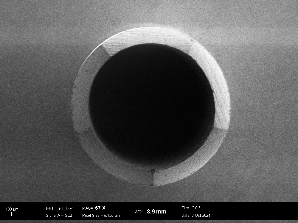
BONE ACCESS NEEDLE™ Cannula – MAG 57X – EHT = 5.00 kV – WD = 8.9 mm – Tilt 0° – Signal A = SE2 – Pixel size = 5.135 µm
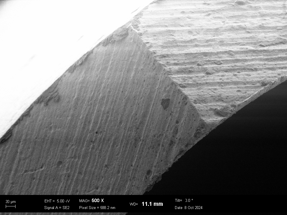
BONE ACCESS NEEDLE™ Cannula – MAG 500X – EHT = 5.00 kV – WD = 11.1 mm – Tilt 0° – Signal A = SE2 – Pixel size = 588.2 nm
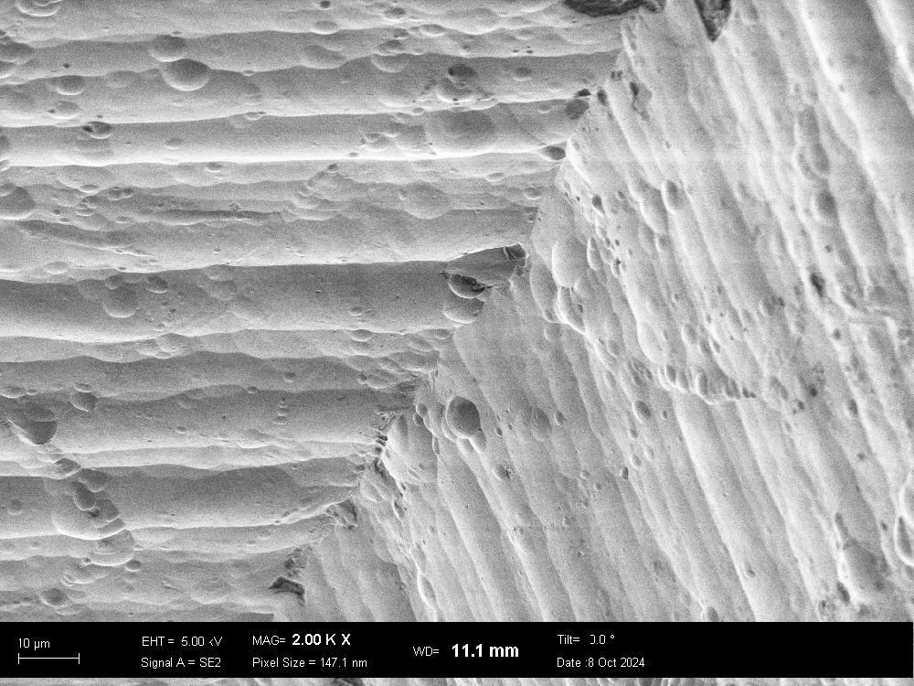
BONE ACCESS NEEDLE™ Cannula – MAG 2.00KX – EHT = 5.00 kV – WD = 11.1 mm – Tilt 0° – Signal A = SE2 – Pixel size = 147.1 nm
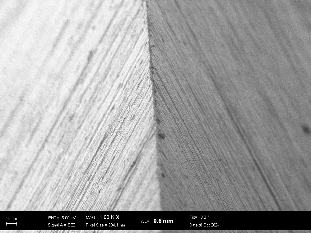
SPEEDYBELL™ INTRODUTTORE – MAG 1.00KX – EHT = 5.00 kV – WD = 9.6 mm – Tilt 0° – Signal A = SE2 – Pixel size = 5.135 µm
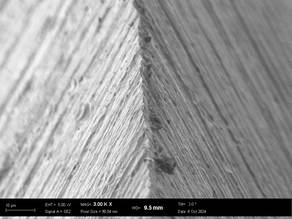
SPEEDYBELL™ INTRODUTTORE – MAG 3.00KX – EHT = 5.00 kV – WD = 9.6 mm – Tilt 0° – Signal A = SE2 – Pixel size = 98.04 nm
SPEEDYBELL™ INTRODUTTORE – MAG 5.00KX – EHT = 5.00 kV – WD = 9.5 mm – Tilt 0° – Signal A = SE2 – Pixel size = 58.82 nm
Understanding SEM Parameters:
To better interpret these images, here are explanations of the key technical terms found in the image descriptions:
EHT (Electron High Tension): The voltage applied to the electron beam, measured in kilovolts (kV). This affects electron penetration depth and image resolution.
Signal A: Indicates the type of signal detected. ‘SE2’ refers to type 2 secondary electrons, which originate from the most superficial layers of the sample and provide high-contrast surface images.
MAG (Magnification): Represents the level of magnification used, e.g., 1.00KX means the image is magnified 1,000 times.
Pixel Size: The dimension of a single pixel in the image, measured in nanometers (nm). A value of 294.1 nm means each pixel corresponds to that physical size.
WD (Working Distance): The space between the sample and the microscope’s objective lens.
Tilt: The angle at which the sample was positioned during scanning.
These high-resolution SEM images confirm the superior sharpness of our needle cutting edges, a crucial feature ensuring smoother and less traumatic insertions.
Through precision engineering and rigorous quality control, we continue to enhance patient comfort and procedural efficiency, reaffirming our commitment to innovation in medical device technology.