METAPHYSEAL BONE FRACTURE TREATMENT BY BALLOON TECHNIQUE
HOME
>
SURGICAL PROCEDURES
>
ORTHOBIOLOGICS PROCEDURES
>
METAPHYSEAL BONE FRACTURE TREATMENT BY BALLOON TECHNIQUE
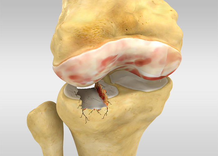
Metaphyseal depression fractures, stemming from trauma or conditions like osteoporosis, pose complex challenges. A prominent example is the tibial plateau fracture, although this procedure applies to proximal humerus, heel, and distal radius fractures as well.
OSTEOPLASTY™ emerges as a groundbreaking solution, facilitating minimally invasive procedures under fluoroscopic guidance.
This technique involves a small balloon catheter that lifts and reshapes bone, complemented by a hardening, radio-opaque, and bioresorbable cement solidifying exclusively in moist environments. Emphasizing percutaneous access, it prioritizes safeguarding the metaphysis and its vascularisation for enduring stability, and it ensures controlled elevation, tissue preservation, swift recovery, and minimal infection risk.
SURGICAL PROCEDURE
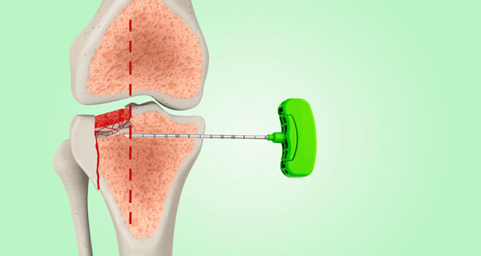

2.
A drill is then inserted to create access for the balloon catheter.
3.
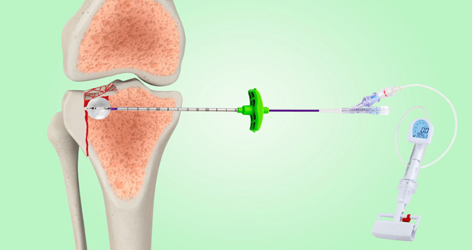
The balloon catheter, loaded with a mix of saline and contrast medium to ensure better visibility during inflation, is inserted through the Working cannula and inflated until the desired physiological anatomy is restored.
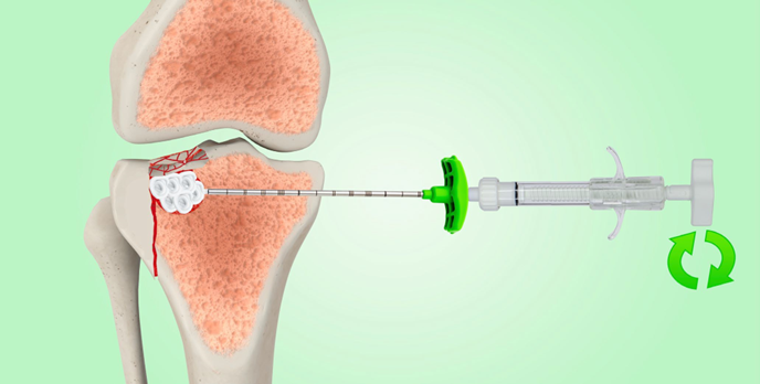
Once deflated and removed the balloon catheter, biological cement is injected in two possible ways: directly into the Luer-lock of the working cannula (OPTION A) or by filling 2.5 ml syringes and bone filler provided in the kit (OPTION B).
4.
Cement injection OPTION A:
Ready-to-use biological cement syringe
The cement syringe is connected to the Luer-lock connection of the working cannula and the cement is released into the area to be treated.

5.
Cement injection OPTION B:
Bone filler cannulas and 2,5 ml syringes
Bone fillers are filled with biological cement and inserted into the working cannula to release cement into the cavity thanks to their plunger.
The lesion should be filled starting from its deepest point, then the bone filler is progressively pulled back to fill it completely.
Empty bone filler before removing it from the working cannula to avoid any leakage, then remove the working cannula.
Subsequently, it is possible to implant screws/plates when necessary.
An arthroscopic procedure can be performed to check that no intra-articular leakage of the biological cement occurred.
Fill out the form for more information.