Home » ORTHOBIOLOGICS PROCEDURES » SUBCHONDRAL BONE MARROW LESIONS TREATMENT
Addressing subchondral bone marrow lesions (BMLs) and avascular necrosis is paramount within the realm of orthopaedics and musculoskeletal health. Often detected through MRI imaging, these conditions signal an array of underlying issues, from osteoarthritis to traumatic injuries and even certain cancers. Frequently affecting weight-bearing joints, they can inflict pain and hinder mobility.
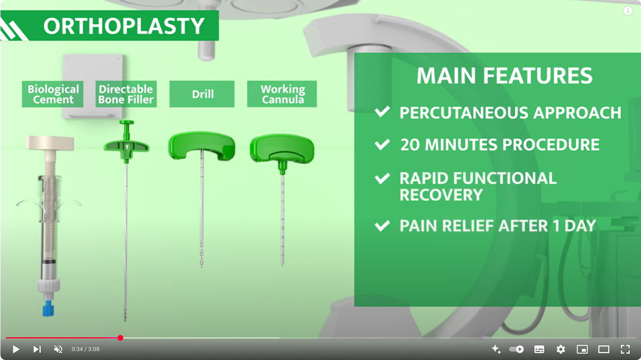
ORTHOPLASTY™ is an innovative surgical toolkit specially designed to combat subchondral bone marrow oedema.
Employing a minimally invasive, percutaneous approach akin to vertebroplasty, this toolkit enables the precise delivery of autologous bone marrow or cutting-edge synthetic biomaterials.
This method significantly reinforces subchondral bone, offering several advantages such as reduced infection risk, rapid functional recovery, pain alleviation within a few days, and the presentation of anatomical integrity across various joints for future interventions. The provided synthetic material is biologic, radio-opaque, and bioresorbable, hardening solely in wet environments. Its ultimate compressive strength surpasses that of healthy cancellous bone, ensuring durability.


Fill out the form below, one of our expert will get in touch with you shortly!
The content of this website is intended exclusively for healthcare professionals.
In accordance with current regulations (Legislative Decree 46/97 and EU Regulation 2017/745), access to information related to medical devices is restricted to healthcare professionals only.
I declare that I am a healthcare professional.