PERCUTANEOUS DISCECTOMY
HOME
>
SURGICAL PROCEDURES
>
SPINE SURGERY PROCEDURES
>
PERCUTANEOUS DISCECTOMY
A hernia is a common disorder that occurs when the intervertebral discs nucleus pulposus becomes dislodged from its natural position inside the annulus fibrosus. Most annulus fibrosus lesions are the result of repeated microtraumas or a major trauma that degenerates to allow the nucleus pulposus to bulge and compress a nerve and/or its surrounding tissues, causing pain in the back and the legs. When conservative treatment fails and symptoms persist or worsen, surgical treatment is considered.
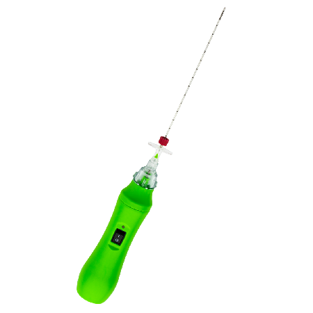
With DISKOM™, pressure on nerve roots or surrounding tissues can be significantly reduced by removing disc herniation through a minimally invasive procedure.
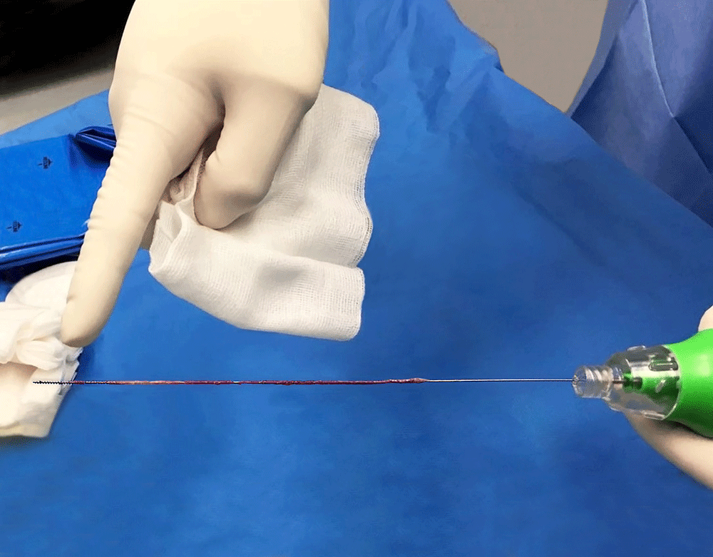
DISKOM™ is made up of two elements: an access needle with both distal and side openings, and a titanium cochlear tip for the mechanical removal of the nucleus pulposus. The great advantage of this device is that the cochlea is tightly connected, so it is almost impossible for it to detach: this is an advantage that is surely a unique feature of this system.
It is a mechanical removal procedure. It does not make use of either radiofrequency or laser. The wide side opening enables the removal of up to 2cc of disc material. The basic principle uses an Archimedes’ screw or cochlea. The Archimedes’ screw works best with fluid or granular material. If the disk is completely dehydrated (black disk), this principle cannot be applied. Totally extruded hernia is also a contraindication.
PERCUTANEOUS DISCECTOMY BENEFITS WITH DISKOM™:
- Annular integrity preservation
- Total procedure time: 10 to 15 minutes
- Up to 2 cc of disc material removed
- Suitable for a bioptic procedure
- Available for thoracolumbar and cervical spine
- Totally single-use device
- No further equipment needed
- No capital investment required
- No thermal damage to the nerve roots
- Percutaneous access → totally MIS procedure
- Fast recovery after treatment
- Just local anaesthesia needed
- Fast discharge of the patient

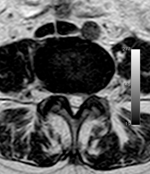
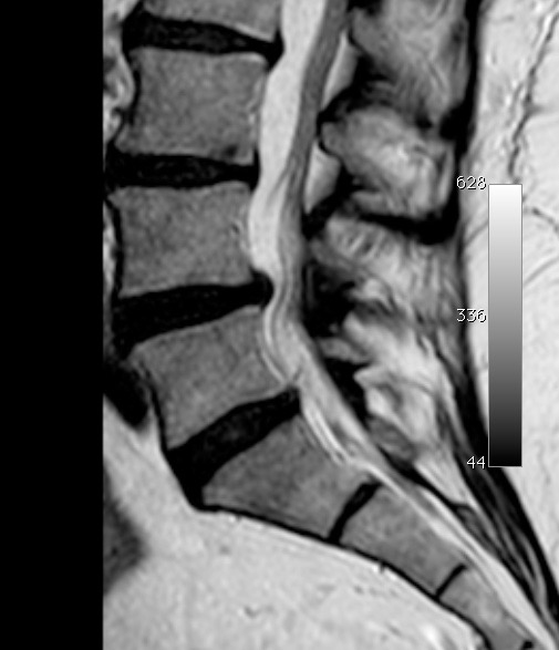
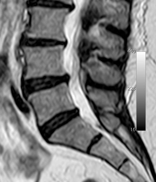
PATIENT SELECTION
- Radicular pain
- MRI consistent with contained disc herniation
- MRI demonstrating 50% preserved disc height
- Failed conservative treatment
- Facet pain excluded
- Positive low-volume diagnostic selective nerve root block
- Discogram and post-disco CT consistent with the above (optional)
SURGICAL PROCEDURE:
- Introduce the access needle through fluoroscopic guidance into the disc. Contrast liquid might be injected through the cannula.
- Remove the stylet and introduce the probe into the cannula, then connect the collection chamber to the access needle cannula, through the Luer-lock connection.
- Switch on the probe.
- Perform a backward and frontward movement, for 2/3 minutes, combining it with a rotational movement.
- The nucleus is removed and collected in the collection chamber or along the probe stylet.
- Remove the device.
RELATED PRODUCT
Discover more about DISKOM™ PERCUTANEOUS DISCECTOMY PROBE:
VIDEO TUTORIAL
Fill the form to request DISKOM™ Case study and Surgical technique.